Skin Cancer
Australia has the highest skin cancer rates in the world courtesy of our largely fairer skinned ancestry, combined with our climate and lifestyle. While perhaps good for the soul, the sun and its ultraviolet radiation is harsh on the body, producing not only premature ageing, but high rates of all types of skin malignancies (cancers).
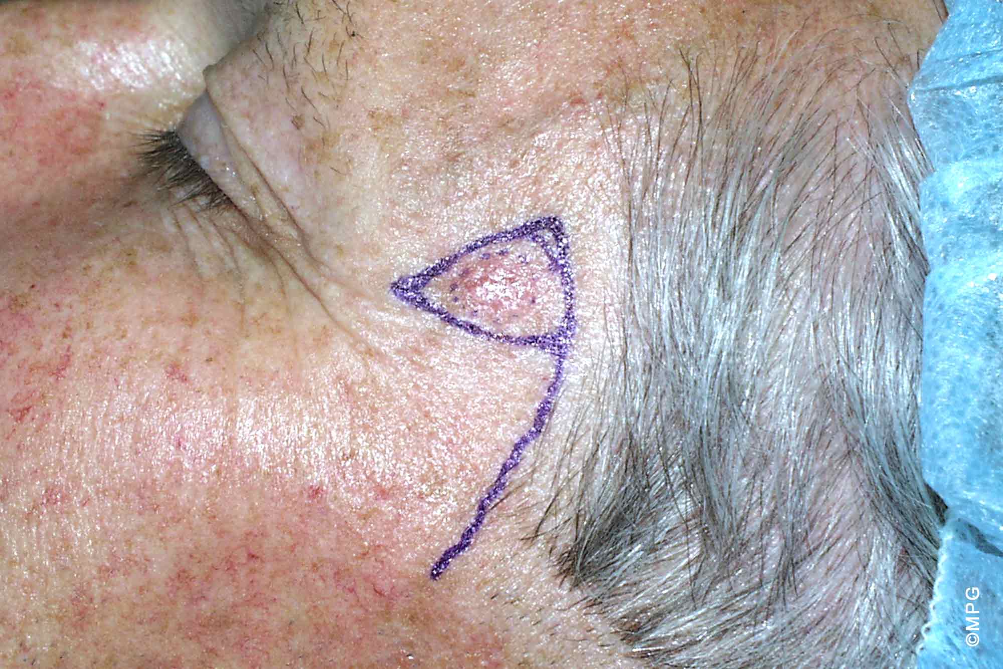
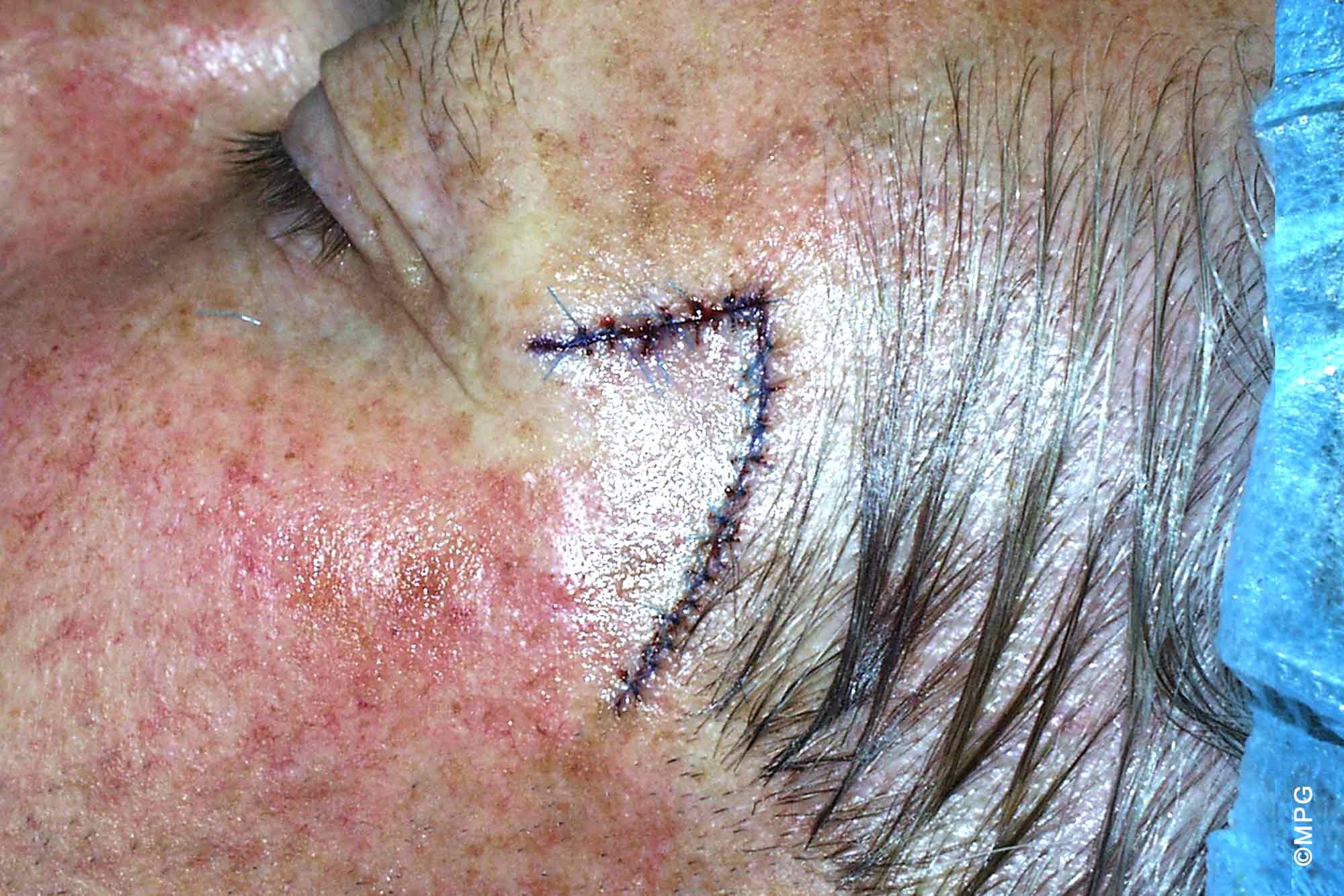
Skin cancers are seen all over the body, but the most common region for the most often seen types is the head, face and neck. These are obviously critical areas that demand experienced and specialised care and knowledge to get the optimal result, not only form the point of view of completely excising the cancer, but also in providing the best cosmetic result from any repair.
There are three main groups of skin cancers:
- Basal cell skin cancers (BCC) – the most common type and generally the least dangerous. In certain sites on the body and certain subtypes of BCC they can be very dangerous and demand respect and experience in their treatment. There are a number of different subtypes of BCC, which behave in different ways with some much more aggressive and ‘infiltrative’ than others. These different types need to be recognised and treated in the manner appropriate for their individual behaviour.
- Squamous Cell Skin Cancers (SCC) – while less common than BCC they are still very frequently seen. They tend to at least have the potential to behave in a more aggressive and malignant manner, with the prospect of spread to lymph glands and other parts of the body.
- Malignant melanoma – this particularly malignant and dangerous form of skin cancer is seen frequently in this country. It can occur in any part of the body – even those parts not usually exposed directly to the sun. Around half of the cases of melanoma develop in existing moles or naevi. The key to melanoma treatment, apart from preventing undue sun exposure, is early diagnosis. The features to look for include:
A – Asymmetry or irregularity of shape
B – Borders that are irregular or ragged
C – Colour, there is often variation in the shade of brown, black or even loss of pigmentation
D – Diameter, harmless moles are usually less than 6mm in diameter.

Our Procedures
Our philosophy is to treat all patients as we would be expected to be treated ourselves.
A/Prof. Mark Gianoutsos